Diopsys® ffERG/Flash Plus Photopic Negative Response Launches to Eye Care Practices Globally
Diopsys Inc, the world leader in modern visual electrophysiology, announces the release of the Diopsys® ffERG/Flash Plus Photopic Negative Response vision test, a new full field electroretinography (ffERG) protocol designed to detect early signs of glaucoma.1-6 By 2050, it is estimated that 6.3 million Americans will have glaucoma, with black Americans having the highest prevalence rate. This disease can lead to irreversible vision loss and blindness, but with timely treatment, disease progression can be slowed. Since there is no cure for glaucoma, early detection is a key factor in preventing loss of sight.
No single test detects glaucoma, as the disease is diagnosed through a comprehensive eye exam that includes several different tests to help build a clinical picture. Unfortunately, sensitive indicators of early glaucoma are difficult to discern, and results from various tests can be equivocal or unreliable.
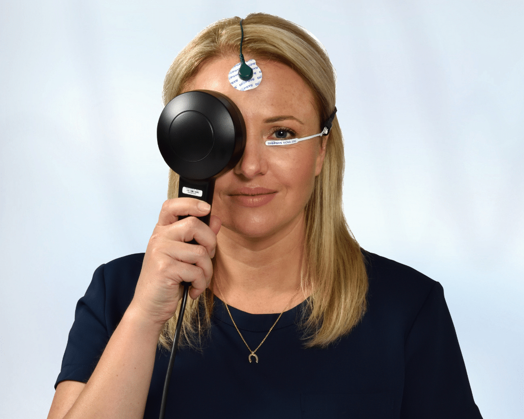
The new Diopsys® ffERG / Flash Plus Photopic Negative Response (PhNR) vision test fills a diagnostic need by providing doctors objective information on the function of retinal ganglion cells (RGCs) and their axons.1 RGCs are the neurons that link the eye to the brain and are the cells primarily damaged by glaucoma. Through evaluating how these cells are functioning, Diopsys® ffERG / Flash Plus PhNR results can provide information on “stressed” cells at a subclinical stage, when they have become dysfunctional but are still alive. Eye care professionals can then begin treatment at this early stage, slowing the progression of the disease, and preserving sight for their patients.
“We are committed to helping the eye care community diagnose glaucoma as early as possible, ” said Alberto González García, MD, Chief Medical Officer at Diopsys. “This new ffERG test provides clinicians with a way to objectively evaluate RGC function efficiently in their practices to make the best clinical decisions for their patients.”
Diopsys also offers another objective test of RGC function called pattern electroretinography (PERG). PERG has been shown to detect RGC dysfunction up to 8 years before changes in RNFL thickness as shown on OCT.7 Both PERG and ffERG/Flash Plus PhNR tests are clinically suitable to evaluate retinal ganglion cell dysfunction and can help eye care professionals in the decision to initiate treatment. The new Diopsys® ffERG / Flash Plus PhNR test has several testing advantages, including the ability to test in a lit room with no refraction needed.
Diopsys plans to showcase Diopsys® ffERG / Flash Plus Photopic Negative Response at several upcoming eye care conferences. For more information, visit Diopsys.com/PhNR.
1. Frishman L, et al. ISCEV extended protocol for the photopic negative response (PhNR) of the full-field electroretinogram. Doc Ophthalmol. 2018;136:207-211. 2. North RV, et al. Electrophysiological Evidence of Early Functional Damage in Glaucoma and Ocular Hypertension. Invest. Ophthalmol. Vis. Sci. 2010;51(2):1216-1222. 3. Viswanathan S, et al. The Photopic Negative Response of the Flash Electroretinogram in Primary Open Angle Glaucoma. Invest. Ophthalmol. Vis. Sci. 2001;42(2):514-522. 4. Machida S, et al. Photopic negative response of focal electoretinograms in glaucomatous eyes. Invest Ophthalmol Vis Sci. 2008;49:5636-44. 5. Gotoh Y, et al. Selective loss of the photopic negative response in patients with optic nerve atrophy. Arch Ophthalmol. 2004;122:341–346. 6. Miyata K, et al. Reduction of oscillatory potentials and photopic negative response in patients with autosomal dominant optic atrophy with OPA1 mutations. Invest Ophthalmol Vis Sci. 2007;48:820–824. 7. Banitt MR, et al. Progressive Loss of Retinal Ganglion Cell Function Precedes Structural Loss by Several Years in Glaucoma Suspects. Invest. Ophthalmol. Vis. Sci. 2013;54(3):2346-2352.


